
⚠ Important Information
This content is for general information only and is not medical advice. For any decision about your health, consult a qualified healthcare professional.
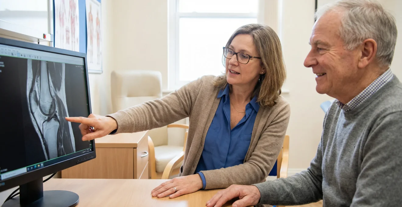
Last week, I sat with Hannah after her orthopaedic appointment. She’d twisted her knee during football, and the MRI report mentioned “meniscal signal change.” Her physio said one thing, the scan report said another, and she left more confused than when she arrived. Sound familiar? Here’s what changed everything: when they did a proper hands-on exam plus a weight-bearing X-ray, the whole picture shifted. Suddenly, the plan moved from “maybe surgery” to “structured rehab with clear triggers for review.”
The NHS performed 96,000 knee replacement procedures in 2023/24. Yet for every person who ends up in surgery, dozens more sit in waiting rooms clutching MRI reports, wondering if that “abnormal finding” means they’re headed for the operating theatre. The reality? Your scan results are just one piece of a much larger puzzle.
The 30-second version: 6 things that actually drive the decision
- Your symptoms and function matter more than any single scan result
- Weight-bearing X-rays often reveal more about arthritis than expensive MRIs
- A proper hands-on exam can be as accurate as imaging for ACL tears
- “Abnormal” findings on MRI are common in pain-free knees
- Your activity goals (sport vs daily function) change the surgical equation
- Red flags like a locked knee need urgent assessment regardless of imaging
You’re looking at your knee scan results right now, aren’t you? Maybe the report uses words like “degeneration” or “tear” and you’re thinking surgery is inevitable. Let me stop you right there. In the consultation stories I’ve reviewed from UK outpatient settings, the classic trap is letting the MRI report make the decision. This observation is limited to the contexts I’ve seen; it can vary with the examiner’s skill and what you actually want (return to sport vs pain-free stairs).
The gap between what imaging shows and what you feel can be massive. I’ve seen marathon runners with “terrible” MRIs who run pain-free, and I’ve reviewed cases where “minor” scan findings masked significant instability that only showed up during physical testing. The disconnect happens because imaging captures structure, not function. It’s like judging whether a car runs by looking at a photograph – you need to turn the key and check under the bonnet.
Your scan is not a verdict: what a knee ‘surgical workup’ really is
When I review knee consultation summaries, the patients who get clear answers fastest aren’t those with the most scans. They’re the ones whose clinicians consider four elements together: the injury mechanism (how it happened), current symptoms (what you feel), physical examination (what the knee actually does), and then – only then – the imaging results.
Think about it this way: if you walked into A&E with severe knee pain but a “perfect” MRI from last month, would they send you home? Of course not. They’d examine you, check for swelling, test stability, and make a decision based on what’s happening now, not what a machine saw weeks ago. The surgical workup works the same way – it’s about connecting dots, not collecting scans.
Here’s what throws people: we’ve been trained to think technology trumps everything else. More pixels, stronger magnets, clearer pictures – surely that’s the answer? But knee surgery decisions aren’t made by radiologists reading screens in dark rooms. They’re made by surgeons who’ve examined thousands of knees, felt the difference between a tight ACL and a loose one, and know that a patient’s goals matter as much as their anatomy.
The hierarchy actually runs opposite to what most people expect. First comes your story – sudden twist with a pop versus gradual onset over months tells us different things. Next is what you can and can’t do – giving way on stairs versus aching after a long walk points to different problems. Then comes the physical exam – the tests that make your knee move in ways that reveal what’s wrong. Imaging comes last, to confirm suspicions or rule out surprises, not to make the decision alone.
Imaging that changes the plan (and imaging that mostly doesn’t)
Let me share something that surprises most patients: that expensive MRI you’re waiting weeks for might tell us less about your arthritis than a simple standing X-ray. According to NICE guidelines for knee osteoarthritis imaging, imaging should only be used when atypical features suggest an alternative diagnosis. Yet patients arrive clutching MRI reports, convinced these determine everything.
The confusion partly stems from how we talk about different scans. Everyone “knows” MRI is more advanced, so it must be better for everything, right? Wrong. Each imaging type answers specific questions, and ordering the wrong test is like using a microscope to read a map – impressive technology applied to the wrong problem. When an orthopaedic knee surgeon evaluates your imaging, they’re matching the scan type to the clinical question, not just ordering the most expensive option available.
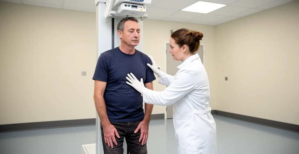
Weight-bearing X-ray: the fastest way to understand arthritis and alignment
You’re standing in the imaging department, and they ask you to put your full weight on that painful knee for an X-ray. Seems cruel, doesn’t it? But here’s why it matters: arthritis behaves differently under load. That joint space that looks fine when you’re lying down for an MRI? It might completely disappear when you’re standing. The mechanical axis – whether your leg bears weight straight or at an angle – only shows up properly when gravity’s involved.
Weight-bearing views reveal what surgeons call the “functional truth” of your knee. If you’ve got bone-on-bone contact happening every time you stand, that’s surgical territory. But if the joint space maintains itself under load, even with some narrowing, you might have years of non-surgical options ahead. This distinction drives the choice between high tibial osteotomy (realigning the leg), partial knee replacement (one compartment), or total knee replacement (whole joint).
I’ve reviewed cases where patients spent hundreds on private MRIs showing “severe degeneration,” only to have a standing X-ray show preserved joint space and good alignment. The surgeon’s recommendation? Physiotherapy and weight management, not surgery. The MRI wasn’t wrong – it just answered a question that wasn’t being asked.
MRI: best for ligaments and meniscus, worst for ‘incidental panic’
MRI excels at showing soft tissues – your ACL, menisci, cartilage surfaces. It can spot a complete ACL rupture, map out a bucket-handle meniscus tear, or reveal hidden bone bruising from recent trauma. When symptoms match these findings, MRI becomes invaluable for surgical planning. The surgeon needs to know if that ACL is partially or completely torn, whether the meniscus is repairable or needs trimming, if other ligaments are involved.
But here’s the catch that generates unnecessary anxiety: MRI sees everything, including changes that don’t cause symptoms. Studies show significant percentages of people walking around pain-free have “abnormal” MRI findings. These incidental findings – meniscal signal changes, mild cartilage thinning, tiny cysts – get reported because radiologists must document what they see. But documenting doesn’t mean operating.
Watch for loaded language in MRI reports. Words like “degeneration,” “tear,” “abnormal signal” sound alarming but might describe normal aging changes. I’ve seen patients convinced they need surgery because the report mentions a “meniscal tear,” not realizing their actual pain comes from patellofemoral irritation that doesn’t even show on the scan. The correlation between MRI findings and symptoms can be surprisingly weak, which is why surgeons insist on examining you, not just your images. This brings us to why understanding the value of a full-body MRI scan versus targeted imaging becomes crucial for medical decision-making.
CT and ultrasound: niche tools that become decisive in specific scenarios
CT scanning might seem outdated for knees – after all, it uses radiation and “only” shows bone. But when surgical planning requires precise bone measurements, understanding rotation, or evaluating previous implants, CT becomes irreplaceable. Surgeons use it for complex revision surgeries, assessing bone loss before reconstruction, or planning patient-specific implants. It’s not about diagnosis anymore; it’s about millimeter-precise surgical blueprints.
Ultrasound, often forgotten in knee discussions, shines for specific questions. It shows tendons in motion, guides needle placement for injections, and evaluates superficial structures like the patellar tendon or collateral ligaments. In the right hands, ultrasound can diagnose a quadriceps tear or confirm a Baker’s cyst faster and cheaper than MRI. The limitation? It’s operator-dependent and can’t see deep inside the joint.
Neither CT nor ultrasound gets ordered routinely. They’re problem-solving tools for specific scenarios. If your surgeon orders one, ask what specific question they’re trying to answer – it usually means they’ve identified something precise that needs investigating, not that they’re fishing for problems.
The comparison between imaging types isn’t about which is “best” – it’s about matching the tool to the clinical question. Let me break this down clearly:
| Imaging Type | Best at showing | Decision-changing use | Common trap | When to question need |
|---|---|---|---|---|
|
Weight-bearing X-ray |
Joint space, alignment, arthritis | Arthritis severity, surgical approach | Missing soft tissue problems | If ordered lying down for arthritis |
|
MRI scan |
Ligaments, meniscus, cartilage | ACL/meniscus surgery planning | Over-reading incidental findings | First test for clear arthritis |
|
CT scan |
Bone detail, implant position | Complex reconstruction planning | Radiation for simple problems | Routine pre-op for standard surgery |
|
Ultrasound |
Tendons, fluid collections | Guided injection placement | Can’t see deep structures | Deep joint investigation needed |
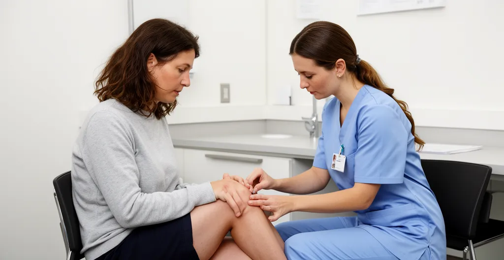
The hands-on exam: clinical tests surgeons rely on (even in the MRI era)
I recently reviewed consultation notes where a patient arrived with a “normal” MRI but couldn’t walk downstairs without the knee buckling. The surgeon spent twenty minutes moving the knee through specific tests, and by the end, the ACL rupture diagnosis was clear. How? Because experienced hands can feel what machines can’t always see – the quality of an endpoint, the subtle give of damaged tissue, the protective muscle guarding that reveals underlying instability.
Clinical tests aren’t random knee wiggling. Each maneuver targets specific structures and problems. The Lachman test checks your ACL by pulling the shin forward with the knee slightly bent. The pivot shift reproduces that awful giving-way sensation if the ACL is gone. McMurray’s test makes the meniscus complain if it’s torn. These tests have names because they work – when done properly.
Here’s what patients rarely grasp until they experience it: these tests can be remarkably accurate. According to a systematic review of ACL test accuracy, the pivot shift test has 94% specificity – meaning if it’s positive, you almost certainly have an ACL tear. The Lachman test shows 81% sensitivity, catching most complete tears. These aren’t perfect, but in experienced hands, they’re often as reliable as imaging.
The real skill lies in synthesizing multiple tests. A knee with a positive Lachman, positive pivot shift, and appropriate injury history (heard a pop, immediate swelling) has an ACL tear until proven otherwise – even if the MRI is pending. Conversely, rock-solid stability on examination makes a complete ACL rupture unlikely, regardless of what shadows appear on the scan.
Why does examiner experience matter so much? Because these tests rely on subtle feelings – the difference between normal ligament stretch and abnormal looseness, between muscle guarding and true mechanical limitation. I’ve watched experienced physiotherapists catch instabilities that junior doctors missed, simply through thousands of repetitions developing that tactile library. It’s like a mechanic who can diagnose engine problems by sound – experience teaches patterns that textbooks can’t fully convey.
The examination also reveals compensation patterns. Someone with chronic ACL deficiency develops characteristic quadriceps weakness and movement strategies. The way you unconsciously protect the knee during testing tells its own story. These functional adaptations don’t show on any scan but profoundly influence surgical decisions. Can you cope without an ACL using muscle control? Are you a good candidate for rehabilitation first?
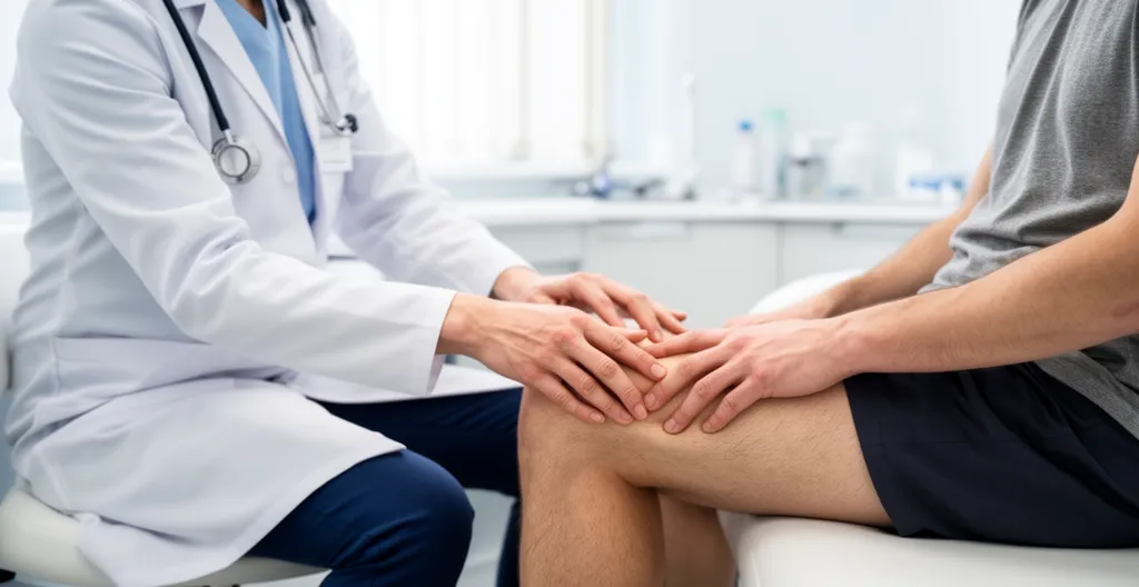
Joint line tenderness deserves special mention. When a surgeon presses along the gap between your thighbone and shinbone, they’re feeling for the meniscus. Precise tenderness at the joint line, especially with a positive McMurray’s test, strongly suggests meniscal pathology. But diffuse tenderness everywhere? That’s often not surgical – it might be widespread inflammation or sensitization that surgery won’t fix.
Watch what happens when examination findings clash with imaging. Stable knee on testing but MRI shows partial ACL tear? Function trumps imaging – if it’s stable, it’s stable. Conversely, clear instability on examination but “intact” ACL on MRI? The surgeon trusts their hands and might order different imaging angles or proceed based on clinical findings. This isn’t arrogance; it’s recognition that functional testing reflects real-world knee behavior better than static pictures.
How findings map to common knee surgeries (and when they don’t)
Let’s cut through the noise: having a “tear” or “degeneration” on your scan doesn’t automatically mean you need surgery. I see this misunderstanding constantly. The decision framework actually works like this: symptoms that limit your life, plus examination findings that explain those symptoms, plus imaging that confirms the diagnosis, plus failure of appropriate non-surgical treatment. Miss any element, and surgery might not be the answer.

ACL and meniscus: instability, locking, and the ‘sport goal’ question
Your ACL is torn. The MRI confirms it, the Lachman test is positive, your knee gives way. Surgery, right? Not necessarily. The crucial factor isn’t the tear itself but whether you have functional instability that limits your activities. Office workers who don’t pivot or jump might do brilliantly with strengthening alone. Footballers who need to cut and change direction? Different story entirely.
The conversation changes completely based on your goals. “I want to return to competitive sport” triggers one pathway. “I just want to walk my dog without the knee giving way” triggers another. Age factors in too – not because older people can’t have ACL reconstruction, but because recovery timelines and activity demands differ. A 50-year-old recreational tennis player might choose stability exercises over nine months of surgical rehabilitation.
Meniscus tears follow similar logic but with extra complexity. That “degenerative” tear in your 45-year-old knee? It might have been there for years without causing problems. Unless you have mechanical symptoms – true locking where the knee gets stuck, or catching that physically blocks movement – arthroscopic surgery often disappoints. Studies show trimming degenerative tears without mechanical symptoms rarely beats good physiotherapy.
But traumatic meniscus tears in younger knees? Completely different situation. A bucket-handle tear in a 25-year-old athlete usually needs urgent surgery. The torn piece flips into the joint, mechanically blocking movement. No amount of strengthening fixes mechanical blockage. The examination finding of a locked knee that won’t fully extend trumps everything else – that’s surgical territory.
Arthritis: why weight-bearing X-rays and alignment often decide more than MRI
When arthritis enters the picture, the decision framework shifts entirely. Forget about tears and instabilities – now we’re talking about bone-on-bone contact, alignment, and quality of life. That weight-bearing X-ray showing complete loss of joint space in one compartment tells us more than any MRI about your surgical options.
Alignment drives surgical planning more than severity alone. If your arthritis affects mainly the inner compartment and your leg angles inward (varus), a high tibial osteotomy might buy you a decade. The surgeon cuts and realigns your shinbone to shift weight off the damaged area. But if multiple compartments are shot, or you’re not active enough to justify the recovery, knee replacement makes more sense.
Here’s what surprises people: “bone-on-bone” doesn’t automatically mean immediate surgery. I’ve reviewed cases of patients functioning well despite no visible joint space, while others with “mild” arthritis on X-ray couldn’t sleep from pain. Function and symptoms drive timing, not pictures. The surgeon weighs your pain levels, activity limitations, response to injections, and life circumstances. A teacher who can’t stand through classes has different needs than a retiree who’s uncomfortable but managing.
Patella and anterior knee pain: when ‘normal imaging’ still leads to a clear plan
Front-of-knee pain frustrates everyone because imaging often looks fine. Your MRI shows intact cartilage, no tears, maybe some “mild signal change” – yet stairs feel like torture. This disconnect happens because patellofemoral pain often stems from tracking issues, muscle imbalances, or overload patterns that static imaging can’t capture.
The physical examination becomes paramount here. Does your kneecap track sideways? Is there crepitus (grinding) with movement? Can you do a single-leg squat without the knee diving inward? These functional findings, combined with your history (gradual onset, worse with sitting), often provide clearer direction than any scan.
Surgery for patellofemoral problems follows strict criteria. Recurrent dislocation with proven instability might need reconstruction. Severe cartilage damage in young patients might warrant cartilage procedures. But the vast majority of front-knee pain responds to targeted rehabilitation addressing hip strength, quadriceps balance, and movement patterns. When the exam shows weakness and poor control rather than structural damage, surgery rarely helps.
Understanding how these different scenarios map to surgical decisions helps you navigate consultations more effectively. Let me break down the typical decision pathway:
A practical triage: what usually drives ‘surgery vs rehab first’
-
If you have red flags (locked knee, suspected infection, major trauma):
Urgent clinical assessment needed. These override all other considerations.
-
If you have mechanical instability (giving way) with positive stability tests:
Discuss ligament reconstruction pathway, especially if you need to return to pivoting sports. Consider your activity goals and recovery timeline tolerance.
-
If you have pain with weight-bearing plus X-ray showing arthritis/alignment issues:
Explore arthritis management pathway. Surgery timing depends on symptom severity, response to conservative treatment, and life impact.
-
If you have mostly pain without instability and function is improving:
Structured rehabilitation first with clear review triggers. Surgery only if specific mechanical symptoms develop or function plateaus despite good rehab.
Bring this to your appointment: a short checklist, then the questions people really ask
I talked to someone last week who had three consultations, three different opinions, and still felt lost. The problem? Each appointment covered different ground because they weren’t prepared with the right information. The surgeon spent half the time trying to understand the timeline, the physio never saw the actual images, and nobody discussed what “success” meant to this specific patient.
Getting a clear surgical opinion isn’t about having more scans – it’s about presenting your case clearly and asking the right questions. When clinicians have complete information and understand your goals, recommendations become much clearer. You’re not just a knee; you’re a person with specific needs, fears, and objectives.
Your 2-minute knee consult pack (what to bring, what to track)
-
A one-line injury story: what happened + when
-
Top 3 functional limits (stairs, running, pivoting, sleep)
-
What makes it worse/better (including rehab tried)
-
All imaging (actual images + reports) and prior op notes if any
-
Your goal: return to sport vs daily mobility vs pain control
-
A list of medications, allergies, and key medical history
The single most important thing you can clarify is your goal. “Fix my knee” isn’t specific enough. Do you need to return to competitive sport? Are you happy if you can walk the dog pain-free? Would you accept some discomfort if you could avoid surgery? These aren’t medical questions – they’re life questions only you can answer. But they fundamentally shape whether surgery makes sense for you.
Track your symptoms properly. “It hurts sometimes” tells the surgeon nothing. “Sharp pain on the inner joint line when I twist to the right, worse in the morning, better after walking for 10 minutes” paints a picture. Keep a simple diary for two weeks before your appointment. Note what triggers pain, what helps, and how it affects your daily activities. This real-world data often matters more than scan reports.

Don’t hide previous treatments, even if they “failed.” That cortisone injection that helped for two weeks tells us something different than one that did nothing. The physiotherapy that made things worse suggests different problems than physio that helped partially. Failed treatments aren’t failures – they’re diagnostic information that shapes the next steps.
For international patients or those considering treatment abroad, there’s another layer of complexity. You’re not just choosing a treatment; you’re navigating different healthcare systems, potentially coordinating logistics for a medical evacuation or planned surgical travel. The preparation becomes even more critical when you won’t have easy follow-up access. Make sure your home medical team and destination surgeon communicate directly about post-operative care plans.
Remember that surgical consultations aren’t sales pitches. A good surgeon should explain why surgery might NOT be right for you as clearly as why it might help. If someone guarantees perfect results or pushes urgent surgery without discussing alternatives, get another opinion. The best surgeons know that happy patients are those with realistic expectations who’ve explored all appropriate options.
The doubts people have right before deciding on knee surgery
If my MRI shows a meniscus tear, do I automatically need surgery?
Not at all. Many meniscus tears, especially degenerative ones in people over 35, don’t require surgery unless you have mechanical symptoms like true locking or catching. If your main complaint is pain without mechanical blockage, structured physiotherapy often works as well as surgery.
Why do I need an X-ray if I already have an MRI?
Weight-bearing X-rays show things MRI can’t: how your joint space changes under load and your leg alignment when standing. For arthritis assessment and surgical planning, these standing views often provide more decision-relevant information than an MRI taken lying down.
Can imaging miss an ACL tear (or overcall one)?
Yes to both. MRI can miss partial ACL tears or show false positives from previous injuries that have healed functionally. This is why clinical examination remains crucial – an experienced examiner can often diagnose ACL status as accurately as imaging through specific stability tests.
What are the red flags that should speed up assessment?
Seek urgent assessment for: a truly locked knee that won’t straighten or bend fully, severe swelling with fever (possible infection), inability to bear weight after acute injury, or significant deformity. These symptoms need evaluation within days, not weeks.
When is a second opinion genuinely worth it?
Consider a second opinion when: surgery is recommended based solely on imaging without examining you, you’re offered surgery as the only option without trying conservative treatment, the recommended procedure seems excessive for your symptoms, or when your gut feeling says something’s off. Different surgeons may have different thresholds and techniques.
Your next move: turning information into action
You’ve got the knowledge now. You understand that your scan results don’t dictate your future, that physical examination matters as much as imaging, and that your goals should drive the decision as much as any test result. The question is: what do you do with this information?
First, stop catastrophizing about that MRI report. Those scary words don’t automatically mean surgery. Focus instead on function – what you can and can’t do, what you want to achieve. If your knee works for your life, abnormal imaging might be irrelevant. If it doesn’t work despite “normal” scans, don’t let anyone dismiss your concerns.
Before your next appointment, honestly assess whether you’ve given conservative treatment a proper chance. Structured rehabilitation means 12 weeks minimum of targeted exercises, not a few sessions then giving up. Many people opt for surgery thinking it’s quicker, not realizing that surgical recovery often takes longer than good conservative care would have taken.
Finally, remember that choosing surgery – or choosing not to have surgery – isn’t a permanent decision. You can try conservative treatment and opt for surgery later if needed. You can have surgery and still need to manage expectations about outcomes. The key is making an informed decision based on accurate information, realistic expectations, and clear personal goals.
The next time someone shows you their knee scan and asks, “Do I need surgery?”, you’ll know the answer isn’t in that image. It’s in the story of their injury, the reality of their symptoms, the findings on examination, and most importantly, what they want from their knee. Armed with the right questions and realistic expectations, you’re ready to have the conversation that actually matters.